
HYMS MedTech was setup to educate and inform medical students and academics on the application of medical technology within medicine. We encourage colleagues from all disciplines to come together to have a platform to innovate and improve current medical practice through the use of medical technology. On the 6th November 2018, HYMS MedTech hosted their inaugural talk at the University of Hull. Our speaker was Dr Raghu Lakshminarayan, Interventional Radiologist at Hull Royal Infirmary, and his talk was aimed at educating students about how technology has influenced the evolution of Abdominal Aortic Aneurysm (AAA) repair. Dr Lakshminarayan highlighted the close relationship interventional radiology has with technology and he explored topics of intravascular procedures, technological innovations, and 3D printing.
Dr Lakshminarayan took us through the history and evolution of AAA repair, exploring old redundant techniques, ‘strokes’ of genius and the ‘piercing’ edge of therapeutic technology used in the present and foreseeable future. The talk focussed on the capabilities and triumph in treating AAA as well as providing us an insight into what interventional radiology (IR) is all about. IR itself is hard to define as a speciality; it consists of multiple procedures over many organ systems of the human body. Fundamentally, it is image-guided minimally invasive techniques which provide either diagnostic information or therapeutics via vascular or non-vascular access. (1,2).
IR is a relatively new medical speciality, finding its feet in the early 1960s with Dr Charles Dotter, a radiologist in the USA. His aspiration in medicine was his desire to replace the scalpel with a catheter and hence pioneered percutaneous transluminal angioplasty; and thus, IR was born. Even though the first official procedure was in 1964, it was not widely accepted until 1974 when cardiologist Dr Andreas Grüntzig furthered the technique and wrote a letter to the Lancet.(3) Dr Lakshminarayan noted that IR does not have the same historical prestige as the older specialities like cardiology or anaesthetics and therefore perhaps demanded less ‘respect’. These specialities have been around far longer and have changed healthcare tremendous amounts, whereas IR has only been around for a minimal 40 years. However, IR has the potential to play a big role in the future of medicine via pioneering minimally invasive techniques. IR is used for procedures in other specialities like cardiology, neurology and surgery, and could well shape other specialities’ futures. Interventional radiologists have a pioneering nature fuelled on new technology as we will explore further through the evolution of AAA repair.
The past, present, and near future of Abdominal Aortic Aneurysm repair
The story of grafts being used to repair AAAs starts in 1951. Dr Lakshminarayan explained that Dr Charles Dubost, a surgeon living in France, was the first to perform a resection of a AAA and replace it with a homograft. This shook the surgical community all over the world, inspiring other surgeons to perform the same procedure.(4) Previously, AAA were suggested to be too big to repair until this technique was developed.(5) However, this was quickly surpassed by Polytetrafluoroethylene (PTFE) grafts. PTFE has a wide range of uses, such as wiring aerospace craft and adhesion procedures as well as vascular prosthetics. Dr Lakshminarayan had these materials on hand for his talk allowing us to test the durability of these space – age materials; it was incredible to feel materials which could survive the desolate, cold vacuum of space in our hands. This biomaterial has a high tensile strength and durability to sustain the constant pulse pressure from the heart. It is also chemically inert therefore it does not react with the body and it is configured to allow for micropores, so the body can heal around/through it. With all these properties, it has become a mainstay in AAA repair.(6–8)
In 1991, Dr Juan Parodi performed the first intravascular repair of AAA. This christened the beginning of repairing AAA endovascularly. During the 1970s, Parodi had the initial idea of repairing AAA endovascularly, however, he did not have the tools until he met a Dr Palmaz who had been perfecting his own endovascularly balloon-expandable stent design. This design was able to give an aortic wall seal and anchorage suitable for Parodi’s vision. Together, Parodi and Palmaz created the first endovascular aortic repair (EVAR).(9,10) Since the inception of EVAR, there have been a number to studies suggesting there are issues with the long-term durability of the graft. A myriad of problems have arisen such as metal fractures, holes in the graft, and device fatigue which could potentially harm the patient. Hence, with EVAR graft, there needs to be monitoring for the foreseeable future.(11) To tackle this problem many companies have become involved in the field. To my surprise, Dr Lakshminarayan gave the example of the Gore Company. I knew them as an outdoor clothing company supplying the waterproof material Gore-tex, however, he told us they had branched out into the medical field and created their own vascular device called the Gore Excluder in the early 2000s. This is a perfect example of a multidisciplinary team requiring material scientist, biomedical engineers, and health professionals all working in tandem to create a complex and potentially life-saving device.(6,12)
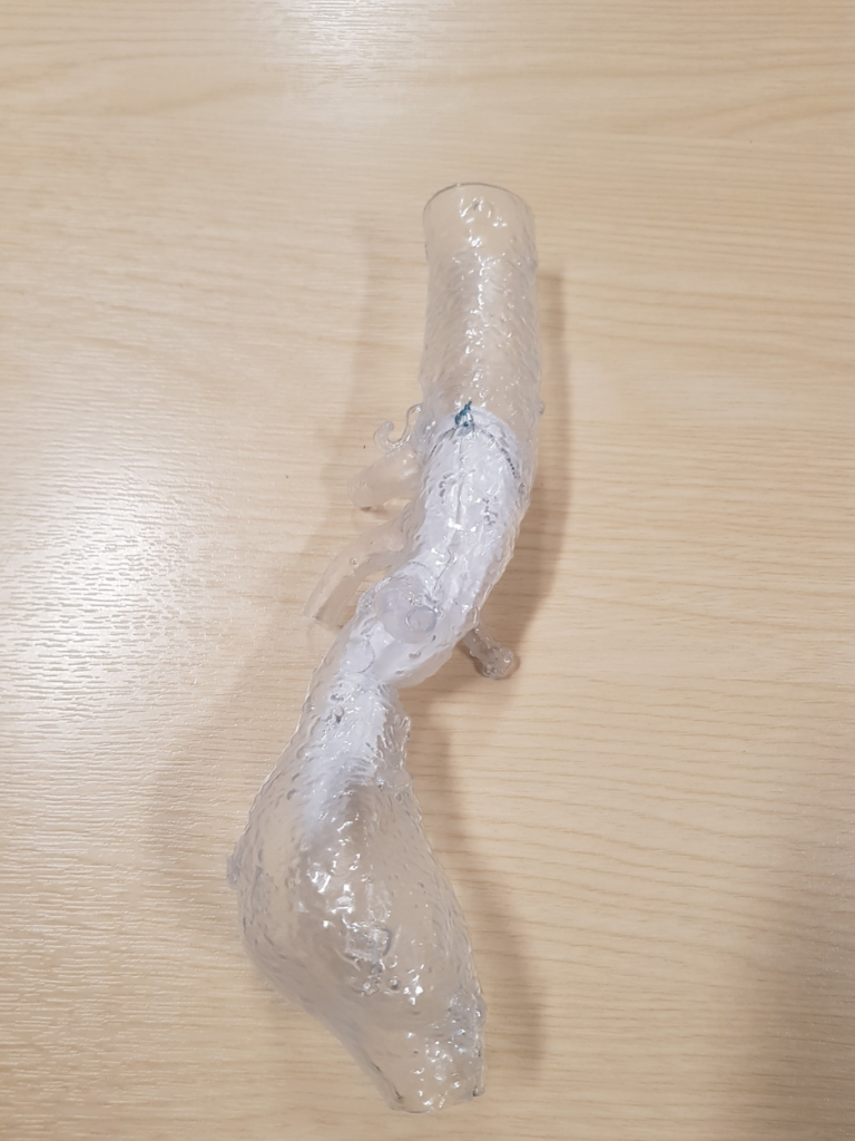
In IR directed AAA repair, there is a major issue which is that anatomy of the patient has hindered its breadth of usage: an aneurysm placed juxtarenally cannot be repaired intravascularly due to the renal arteries. Normally, this is a hard task as the graft may not be able to fit properly due to the branching anatomy and it may be prone to subsequent endoleaks. However, with the advent of 3D printing technologies, Dr Lakshminarayan explain that now we can 3D print a patient’s AAA using images from medical scanners. It simulates the patient’s AAA allowing for personalisation of the graft appropriately to minimise endoleaks, making a tighter and more anchored fit.(13,14) In the case of Hull, images are sent to Glasgow and they create the grafts. When Dr Lakshminarayan was asked what the future could hold, he mentioned that having a pressure transducer in the graft could feed data to the doctor to monitor the aneurysm sac pressure and graft viability.(15,16)
3D printing applications
Dr Lakshminarayan’s talk led up to how 3D models are printed and then used to aid the creation of the personalised graft. Radiology combined with computational modelling has been used a lot in 3D printing as the ability to make detailed 2D images and turn it into a 3D model or prostheses has augmented surgical planning and treatment methods respectively.(17) The ability to ‘print’ any complex shape at a quick pace which is also cost effective, this is what makes 3D printing very viable in the future of medicine. Combined with radiology this makes it the perfect tool to visualise pathology inside the body and bring it outside as a model. I feel 3D printing in the future has great promise in the field of surgery.(18) The most notable use is the possibility of bioprinting complex organs from autologous human cells, this would hopefully cancel out the organ shortage plaguing transplant lists and provide a new hope for patients with organ failure. Bioprinting is the layering of cells and bioinks onto a compatible biomaterial scaffold to create an organ. Unfortunately for now, progress is limited by the complexity of blood vessels within the bioprinted organ.(18)
The future of Interventional Radiology
American computer scientist Alan Kay once said: ‘The best way to predict the future is to invent’. This phrase for me sums up IR. Dr Lakshminarayan‘s talk demonstrated that Interventional Radiology is a pioneer driven speciality as shown previously by Dotter. Dr Ivan Seldinger, a radiologist, pioneered the field of interventional cardiology, he invented the procedure of progressing a catheter into the femoral artery. Dr Pierre Gobin and Dr Jeffery Wensel invented the Mechanical embolus removal in cerebral ischemia (MERCI) Retriever, which could retrieve emboli from a stroke patient to restore blood flow to the affected area. Mechanical stroke thrombectomy has been very effective now that neurologists, neurosurgeons, and stroke physicians are being trained to carry the procedure out due to the increased demand.(19,20) IR is driven by the practising individuals who pioneer their own ‘special interest’ perhaps this will lead to greater across a range of specialties and better outcomes for patients.(21,22)
To end, Dr Lakshminarayan’s talk showed us the past, present and a glimpse of the future for Interventional Radiology. Thank you, Dr Lakshminarayan for giving an incredibly informative first talk for HYMS MedTech. Future talks of HYMS MedTech will include robotics in urological surgery and the applications tissue engineering.
~~~~~~~~~~~~~~~~~~~
James Frost is a fourth year medical student at HYMS and Co-founder of HYMS MedTech. He has keen interests in radiology, minimally invasive procedures and regenerative medicine. In his spare time, he enjoys playing rugby, navigating the ocean floor scuba diving and exploring the animal kingdom.
…
References
1.Renai SA, Belli A-M. A career in interventional radiology. BMJ 2014 Aug 11;349:g5040.
2. Overview of Interventional Practice in the UK | The Royal College of Radiologists [Internet]. [cited 2018 Nov 26]. Available from: https://www.rcr.ac.uk/overview-interventional-practice-uk
3. Payne MM. Charles Theodore Dotter: The Father of Intervention. Tex Heart Inst J. 2001;28(1):28–38.
4. Friedman SG. The 50th anniversary of abdominal aortic reconstruction. Journal of Vascular Surgery. 2001 Apr 1;33(4):895–8.
5. The history of abdominal aortic repair: from Egypt to EVAR [Internet]. Australian Medical Student Journal. [cited 2018 Nov 26]. Available from: http://www.amsj.org/archives/2484
6. Faries PL, Dayal R, Lin S, Trociolla S, Rhee J, Kent KC. Endovascular stent graft selection for the treatment of abdominal aortic aneurysms. J Cardiovasc Surg (Torino). 2005 Feb;46(1):9–17.
7. Dhanumalayan E, Joshi GM. Performance properties and applications of polytetrafluoroethylene (PTFE)—a review. Adv Compos Hybrid Mater. 2018 Jun 1;1(2):247–68.
8. The Properties and Advantages of PTFE [Internet]. AFT Fluorotec. [cited 2018 Nov 26]. Available from: https://www.fluorotec.com/blog/the-properties-and-advantages-of-polytetrafluoroethylene-ptfe/
9. Mauro M, Murphy K, Thomson K, editors. Image-Guided Interventions: with Expert Consult – Online and Print; Expert Radiology Series. 2E ed. 1344 p.
10. Schanzer A, Messina L. Two Decades of Endovascular Abdominal Aortic Aneurysm Repair: Enormous Progress With Serious Lessons Learned. J Am Heart Assoc. 2012 Jun; 1(3): e000075.
11. Jacobs TS, Won J, Gravereaux EC, Faries PL, Morrissey N, Teodorescu VJ, et al. Mechanical failure of prosthetic human implants: A 10-year experience with aortic stent graft devices. Journal of Vascular Surgery. 2003 Jan 1;37(1):16–26.
12. Gore Medical | Vascular grafts, endovascular, interventional, and surgical devices [Internet]. [cited 2018 Nov 26]. Available from: https://www.goremedical.com/
13. Meess KM, Izzo RL, Dryjski ML, Curl RE, Harris LM, Springer M, et al. 3D Printed Abdominal Aortic Aneurysm Phantom for Image Guided Surgical Planning with a Patient Specific Fenestrated Endovascular Graft System. Proc SPIE Int Soc Opt Eng. 2017 Feb 11; 10138: 101380P.
14. Sheth R, Balesh ER, Zhang YS, Hirsch JA, Khademhosseini A, Oklu R. Three-Dimensional Printing: An Enabling Technology for IR. Journal of Vascular and Interventional Radiology. 2016 Jun 1;27(6):859–65.
15. Picel AC, Kansal N. Essentials of Endovascular Abdominal Aortic Aneurysm Repair Imaging: Postprocedure Surveillance and Complications. American Journal of Roentgenology. 2014 Sep 23;203(4):W358–72.
16. Ellozy SH, Carroccio A, Lookstein RA, Minor ME, Sheahan CM, Juta J, et al. First experience in human beings with a permanently implantable intrasac pressure transducer for monitoring endovascular repair of abdominal aortic aneurysms. J Vasc Surg. 2004 Sep;40(3):405–12.
17. Mitsouras D, Liacouras P, Imanzadeh A, Giannopoulos AA, Cai T, Kumamaru KK, et al. Medical 3D Printing for the Radiologist. RadioGraphics. 2015 Nov 1;35(7):1965–88.
18. Ventola CL. Medical Applications for 3D Printing: Current and Projected Uses. P T. 2014 Oct;39(10):704–11.
19. Merci Retriever – Interventional Neuroradiology Specialty – UCLA Interventional Neuroradiology, Los Angeles, CA [Internet]. [cited 2018 Nov 27]. Available from: http://radiology.ucla.edu/merci-retriever
20. Endovascular Today – An Interview With Pierre Gobin, MD [Internet]. Endovascular Today. [cited 2018 Nov 27]. Available from: http://evtoday.com/2005/07/EVT0705_5q.html/
21. Higgs ZCJ, Macafee D a. L, Braithwaite BD, Maxwell-Armstrong CA. The Seldinger technique: 50 years on. Lancet. 2005 Oct 15;366(9494):1407–9.
22. Lakhan SE, Kaplan A, Laird C, Leiter Y. The interventionalism of medicine: interventional radiology, cardiology, and neuroradiology. Int Arch Med. 2009 Sep 9;2:27.